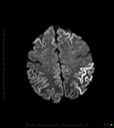
Sporadic Cjd Mri
Diagnosis of sporadic Creutzfeldt-Jakob disease sCJD remains a challenge because of the large variability of the clinical scenario especially in its early stages which may mimic several reversible or. 12 Brain biopsy or autopsy is required for a definitive diagnosis definite CJD.

Creutzfeldt Jakob Disease Radiology Reference Article Radiopaedia Org
More recently a new clinically distinct form of the disease affect.
Sporadic cjd mri. The most frequent is the sCDJ. With respect to sporadic Creutzfeldt-Jakob disease sCJD six molecular subtypes MM1 MM2 MV1 MV2 VV1 and VV2 have been described which vary with respect to age at disease onset disease duration early symptoms and neuropathology. Our literature review yielded a case of NCSE erroneously diagnosed as CJD because similar MRI changes were seen in CJD and peri-ictal states.
MRI lesion profiles in sporadic Creutzfeldt-Jakob disease. Of the four subtypes of CJD described the commonest is sporadic CJD sCJD. MRI signal alterations were reported to correlate with distinct CreutzfeldtJakob disease CJD subtypes.
Diffusion magnetic resonance imaging MRI. It also suggested that MRI. MRI has been shown to be highly sensitive and specific for the diagnosis of sCJD with high inter.
Results The age of onset of E196K genetic CJD cases median of 61 years was older than the E196A cases median of 67 years. Sporadic Creutzfeldt-Jakob disease CJD the most common human prion disease is generally regarded as a spontaneous neurodegenerative illness arising either from a spontaneous PRNP somatic mutation or a stochastic PrP structural change. à propos dun cas MRI role in Creutzfeldt-Jakob disease.
Pan Afr Med J. Probable sporadic CreutzfeldtJakob disease mimicking focal epilepsy. Reporting on the sensitivity and reliability of MRI diagnosis in CJD and the radiological differential diagnosis.
20 were neuropathologically confirmed definite sCJD 19 and 19 were classified as having probable sCJD. Sporadic onset Creutzfeldt-Jacob disease. 3 In sporadic CJD sCJD diagnostic MR examinations are performed frequently and reveal typical findings.
Brain MRI findings are found in most contemporary criteria for the diagnosis of sporadic Creutzfeldt-Jakob disease sCJD including the Centers for Disease Control and Preventions CDC criteria 1. According to WHO diagnostic criteria 1998 identification of 14-3-3 in the CSF is an accurate test to detect sporadic CJD. Meissner B Kallenberg K Sanchez-Juan P et al.
Two Japanese sporadic Creutzfeld-Jakob disease sCJD patients with valine homozygosity at codon 129 of the prion protein gene and protease-resistant prion protein PrPSc type 2 VV2 are described. Sporadic sCDJ familial and acquired. Creutzfeldt-Jakob disease CJD and variant CJD vCJD.
1 Patients were referred to the German CJD Surveillance Unit in the years 20012003 as described previously. It also suggested that MRI ila14-3-3 M might be kMRI may be more useful than the CSF protein 14-3-311. The case was diagnosed as per WHO and MRI-CJD Consortium diagnostic criteria as a probable sCJD during the antemortem stage based on the clinical.
Creutzfeldt-Jakob-Disease CJD a fatal neurodegenerative disorder is diagnosed by the detection of an accumulation of an abnormal form of the human prion protein PrP Sc in the brain. MRI has had an important role in the diagnosis of CreutzfeldtJakob disease CJD. Classification of sporadic Creutzfeldt-Jakob disease based on molecular and phenotypic analysis of 300 subjects.
We studied MR imaging scans of 39 patients with sCJD. MRI of 1458 patients referred to the National Prion Disease Pathology Surveillance Center were collected. The road from first symptoms to diagnosis was a terrifying whirlwind for the whole family lost in the free-fall of being struck by a rare disease.
Crossref Medline Google Scholar. Methods Neurological examination EEG and MRI western blot gene sequence and RT-QuIC. Cerebral biopsy in living patients is to be.
In contrast with Western countries this type of sCJD is very rare in Japan. MRI of Creutzfeldt-Jakob disease. With respect to sporadic CreutzfeldtJakob disease sCJD six molecular subtypes MM1 MM2 MV1 MV2 VV1 and VV2 have been described which vary with respect to age at disease onset disease duration early symptoms and neuropathology.
1 We did not include the cases of patients with possible sCJD. Sporadic iatrogenic recognized risk. According to the WHO diagnostic criteria1 sporadic CreutzfeldtJakob disease sCJD is diagnosed by characteristic electroencephalographic EEG findings the presence of 14-3-3 protein in the cerebrospinal fluid CSF and appropriate clinical symptoms1.
In diffusion weighted n test to detect sporadic CJD. Brain MRI criteria include hypersensitivity on diffusion-weighted imaging DWI or fluid-attenuated inversion recover FLAIR with DWI being the. Imaging of Creutzfeldt-Jakob Disease.
MRI signal alterations were reported to correlate with distinct Creutzfeldt-Jakob disease CJD subtypes. Sporadic CreutzfeldtJakob disease sCJD comprises several subtypes as defined by genetic and prion pro-. Generally these two subtypes of genetic CJD were more like sporadic Creutzfeldt-Jakob disease sCJD clinically.
Creutzfeldt-Jakob Disease CJD is a rare progressive and invariably fatal neurodegenerative disease characterized by specific histopathological features. The introduction of brain magnetic resonance imaging techniques such as fluid-attenuated inversion. Imaging Patterns and.
Sporadic CreutzfeldtJakob disease sCJD is a rare transmissible disease characterized by accumulation of pathologic prion. 2019 Apport de lIRM dans la maladie de Creutzfeldt Jakob. In May 2016 my dad died aged 67 six weeks after being diagnosed with sporadic Creutzfeldt-Jakob disease CJD a rare neurological disorder.
Clinical features are compatible with vCJD and MRI does not show bilateral pulvinar high signal. Magnetic resonance MR imaging summarizing the clinical sce-. Antemortem assessment of sporadic Creutzfeldt-Jakob disease sCJD can be significantly hampered due to its rarity low index of clinical suspicion and its non-specific clinical features.
3 In our study group 19 patients. Three forms of CJD are known. Magnetic resonance Imaging MRI of brain revealed high signal intensity on T2 weighted image T2WI and fluid attenuated inversion recovery sequences in the caudate and putamen bilaterally.
Habibi H et al. On the 19 February 2016 the family had a lovely weekend. The aim of our study was to compare the efficacy of different MRI sequences among six biopsy-proven patients with sporadic CJD sCJD and seven patients with probable sCJD.
Interesting MRI observations. In 123 sCJD cases only two were recognised as VV2 by the Japanese CJD surveillance committee. In our case we cannot conclusively state whether the MRI findings were due to seizure activity or CJD pathology.

Creutzfeldt Jakob Disease Radiology Reference Article Radiopaedia Org

Variant Or Sporadic Creutzfeldt Jakob Disease The Lancet
Fig 2 Pattern Of Cortical Changes In Sporadic Creutzfeldt Jakob Disease American Journal Of Neuroradiology